IB DP Biology: SL復習筆記1.4.4 Skills: Cell Division

Mitotic Phases: Identification
- Cells undergoing different stages of the cell cycle can be identified using photomicrographs taken from microscope slides
- Cells undergoing certain stages of the cell cycle have distinctive appearances
Interphase
- As cells spend the majority of the cell cycle in this stage then most cells will be in this stage
- The?chromatin?is?visible?so the nuclei have a dark appearance
Prophase
- Chromosomes are?visible
- The nuclear envelope is breaking down
Metaphase
- Chromosomes?are lined up along the?middle?of the cell
Anaphase
- Chromosomes are moving away?from the middle of the cell, towards opposite poles
Telophase
- Chromosomes have arrived at?opposite poles?of the cell
- Chromosomes begin to?uncoil?(are no longer condensed)
- The nuclear envelope is reforming
Cytokinesis
- Animal cells: a?cleavage furrow?forms and separates the daughter cells
- Plant cells: a?cell plate?forms at the site of the metaphase plate and expands towards the cell wall of the parent cell, separating the daughter cells
Identification of phases
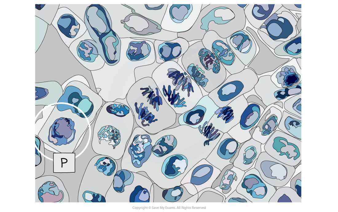
Micrograph showing a cell undergoing prophase (P)
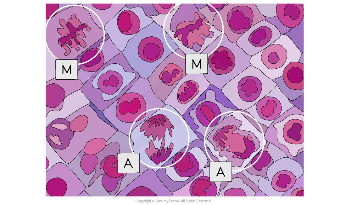
Micrograph showing cells undergoing metaphase (M) and anaphase (A)
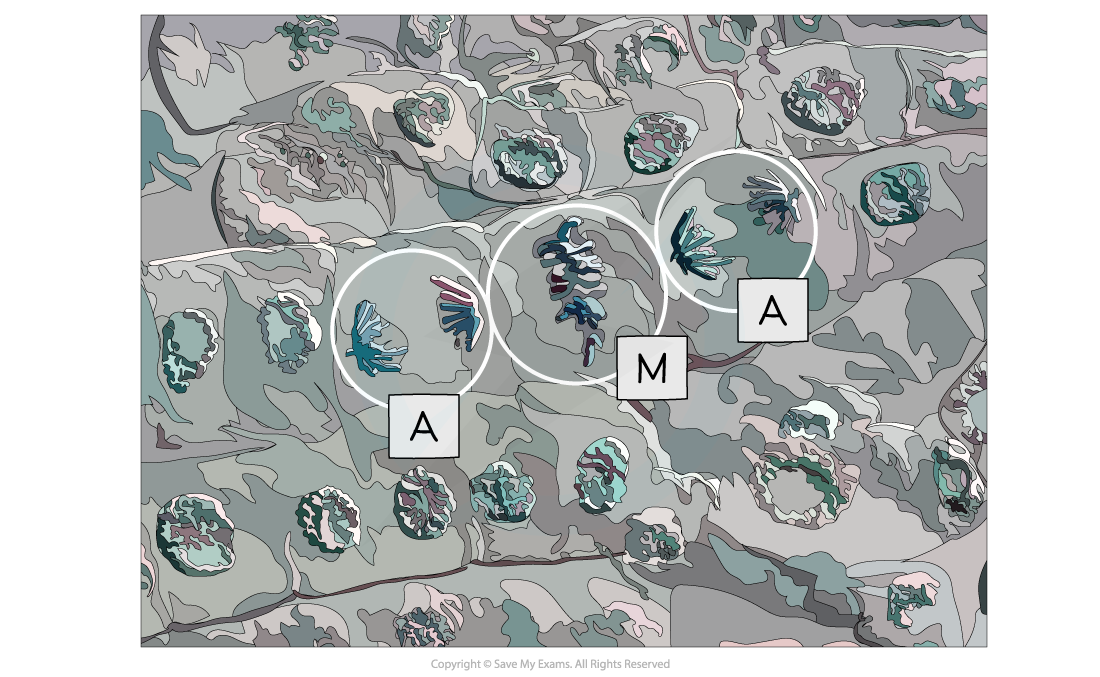
Micrograph showing cells undergoing metaphase (M) and anaphase (A)
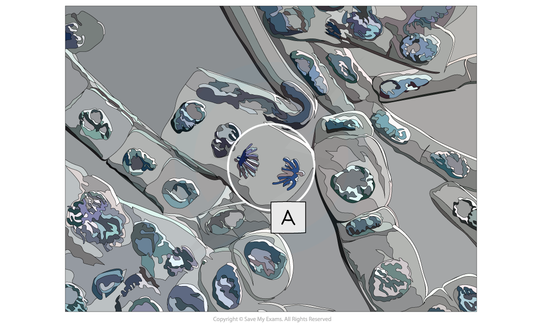
Micrograph showing a cell undergoing anaphase (A)
Exam Tip
It is important to be able to recognise each mitotic stage from electron micrographs and to be able to explain why that cell is in the stage you have selected.It can be difficult to tell prophase and telophase apart in some photomicrographs. In prophase, there is only one group of chromosomes while in telophase there are two groups, one at each pole.
Determination of Mitotic Index
Investigating mitosis in root tissue
- Growth in plants occurs in specific regions called?meristems
- The root tip meristem can be used to study?mitosis
- The root tip meristem can be found?just behind the protective root cap
- In the root tip meristem, there is a?zone of cell division?that contains cells undergoing?mitosis
- Pre-prepared slides of root tips can be studied or temporary slides can be prepared using the?squash technique?(root tips are?stained?and then?gently squashed, spreading the cells out into a thin sheet and allowing individual cells undergoing mitosis to be clearly seen)
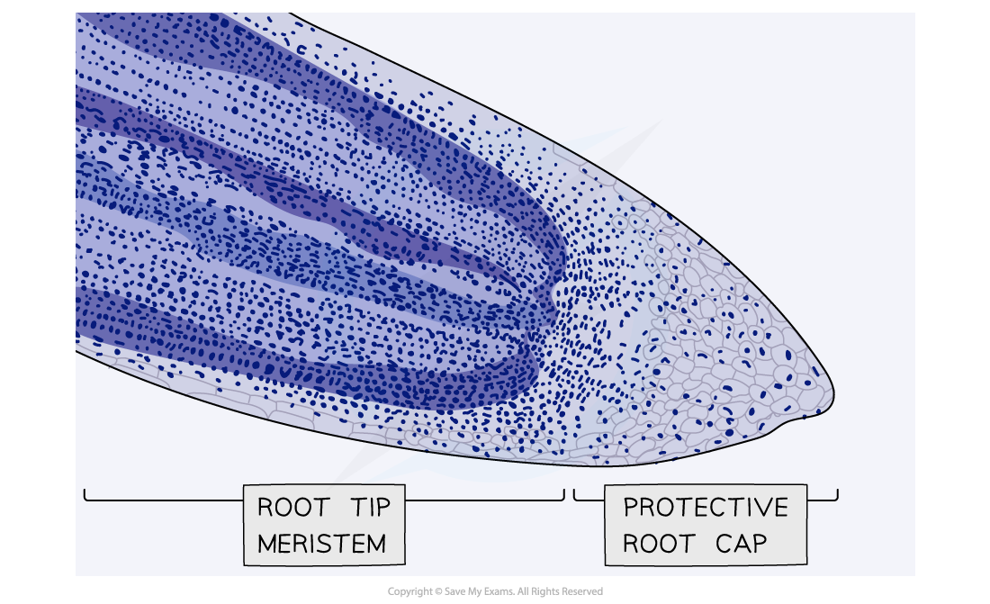
Micrograph showing a stained root tip
Analysis
- Cells undergoing mitosis (similar to those in the images below) can be seen and drawn
- Annotations can then be added to these drawings to show the?different stages of mitosis
- The?mitotic index?can be calculated
The mitotic index
- The mitotic index is the?proportion?of cells (in a group of cells or a sample of tissue) that are?undergoing mitosis
- The mitotic index can be calculated using the formula below:

- You can multiply the answer by?100?if you need to give the mitotic index as a?percentage
Worked Example
A student who wanted to observe mitosis prepared a sample of cells. They counted a?total of 42?cells in their sample,?32 of which had visible chromosomes. Calculate the mitotic index for this sample of cells (give your answer to 2 decimal places).

mitotic index =?0.76
Worked Example
The table below shows the number of cells in different stages of mitosis in a sample from a garlic root tip. Calculate the mitotic index for this tissue (give your answer to 2 decimal places).
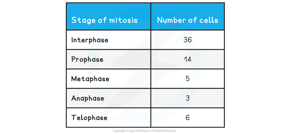



mitotic index =?0.44
Worked Example
The micrograph below shows a sample of cells from an onion root tip. Calculate the mitotic index for this tissue (give your answer to 2 decimal places).
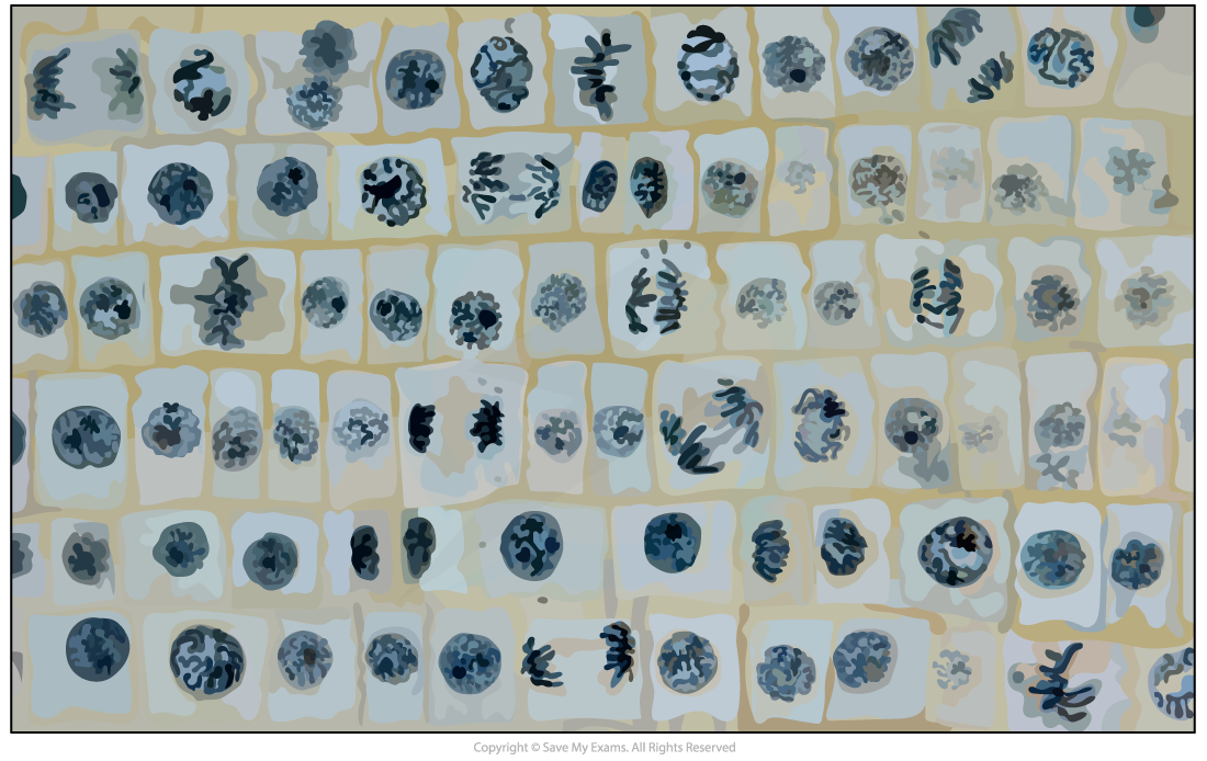
A sample of cells from an onion root tip
Step 1: Identify the cells undergoing mitosis

Number of cells with visible chromosomes (green) = 20
Step 2: Count the total number of cells
Total number of cells (green + red) = 20 + 55 = 75
Step 3: Substitute numbers into the equation

mitotic index =?0.27
Exam Tip
You will need to remember the mitotic index formula as it will not be given to you.
轉載自savemyexams
以上就是關于【IB DP Biology: SL復習筆記1.4.4 Skills: Cell Division】的解答,如需了解學校/賽事/課程動態,可至翰林教育官網獲取更多信息。
往期文章閱讀推薦:
MIT官方發布【2026年夏季推薦閱讀書單】!橫跨科學/人文/經濟...

翰林AMC8視頻課重磅上線!

國際競賽真題資源免費領取